Hatini Lab
Understanding Epithelial Morphogenesis
Tell Me More

What we do?

Morphogenesis is the process that generates the shape of tissues and organs. Our lab seeks to elucidate the molecular and cellular basis of epithelial tissue morphogenesis to better understand basic mechanisms of animal development.

Our Mission

Motivation

Morphogenesis is controlled primarily by local coordinated changes of cell shape, rearrangements of cell-cell contacts, cell proliferation and cell death. The genes and molecular mechanisms that regulate these cellular behaviors are only partially characterized. Therefore, a major goal is to identify novel genes that participate in tissue morphogenesis and elucidate their mechanism of function. Anomalies in epithelial morphogenesis underlie a range of congenital diseases such as spina bifida, cystic kidneys and vascular aneurysm, and acquired diseases such as cancer that disrupt the normal morphology of tissues and organs. To understand these disease processes a deep understanding of the mechanisms involved is essential.

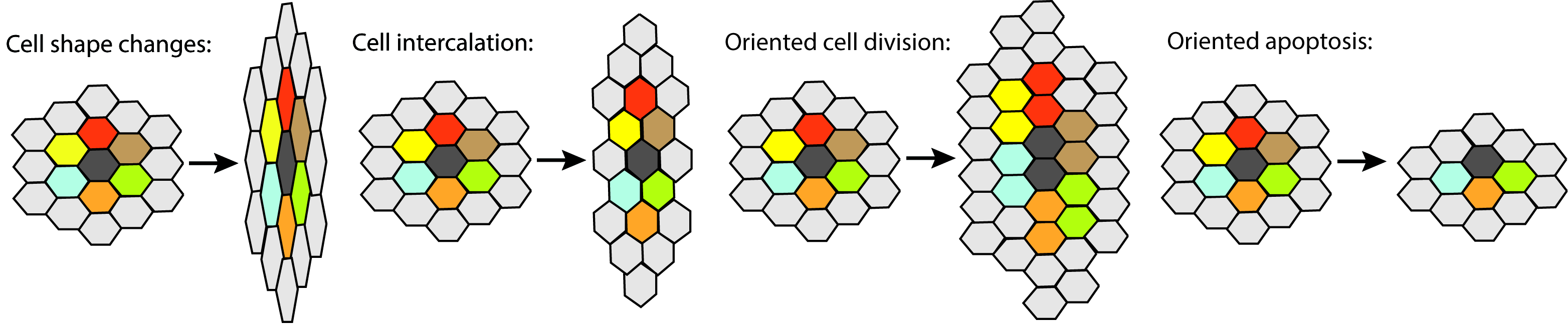
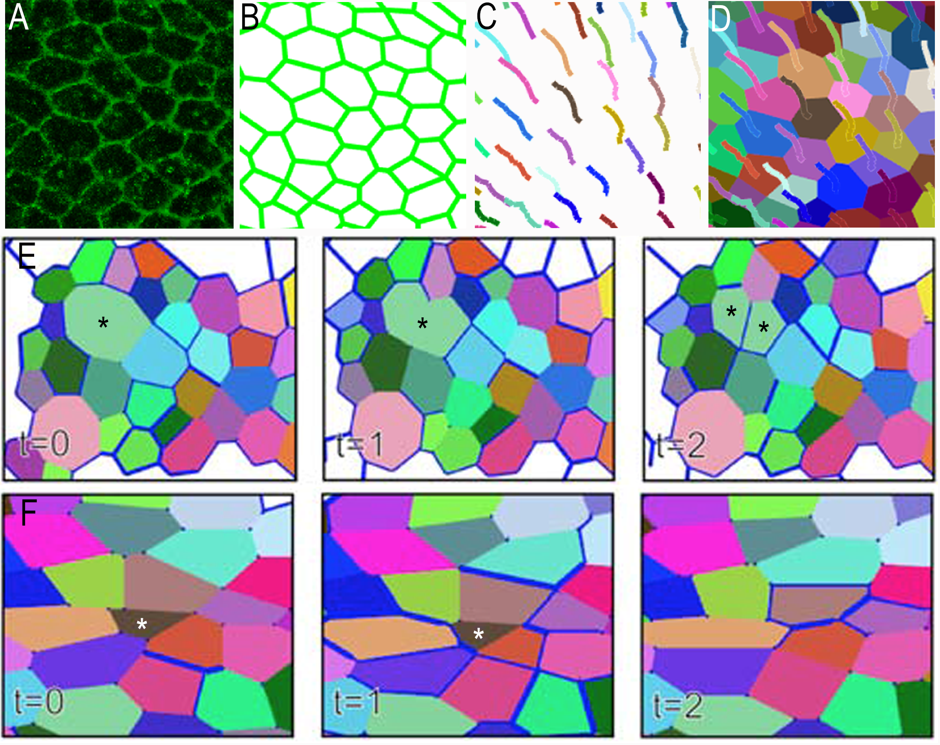
Coordinated cell behaviors drive tissue morphogenesis. Coordinated changes in cell shape, rearrangements of cell-cell contacts, cell proliferation and cell death lead to global changes in tissue morphology. Our lab employs genetic analysis, live imaging and quantitative image analysis to elucidate the molecular and cellular basis of epithelial morphogenesis.

Coordinated cell behaviors drive tissue morphogenesis

Critical barriers to knowledge

Mechanical forces generated by cell proliferation, apoptosis, cell-cell adhesion and the cytoskeleton drive morphogenesis. However, the modulation of mechanical force generation in space and time by extracellular signals, polarized membrane landmarks and cytoskeletal effectors are just being elucidated. The convergence of recent advances in imaging technologies, fluorescent reporters, computational image analysis and mathematical modeling enables developmental biologists to observe and define the molecular and cellular basis of morphogenetic processes with unprecedented detail.

How we do it?

The approaches we use

Our lab investigates the morphogenesis of the epithelium of imaginal discs of fruit fly Drosophila as a model system. The imaginal discs are set aside from the embryonic epidermis during embryonic development. During larval stages, secreted signals called morphogens that emanate from localized signaling centers promote proliferation of epithelial cells and specify the cellular identities of the various parts of adult appendages. During post-larval stages the imaginal discs undergo extensive remodeling to generate the final shape of adult appendages.

Epithelial elongation is a conserved process that alters the proportions of epithelial sheets and tubes. It restructures the early embryo, and tubular epithelia such as the neural tube, the lungs airways, the kidney collecting system, and the collecting ducts of secretory organs. The epithelium of the leg imaginal disc narrows and elongates to form a hollow cylinder during post-larval stages. Our lab employs the leg imaginal disc as a model to investigate the molecular and cellular basis of tissue elongation and the regulation of the process by extracellular signals.

Morphogenesis of the leg imaginal disc is a model for epithelial elongation by cell shape changes and rearrangements of cell-cell contacts.

Morphogenesis of the leg imaginal disc is a model for epithelial elongation by cell shape changes and rearrangements of cell-cell contacts.

The fly eye is composed of ~800 ommatidial units. Shown is a single ommatidium at 28 and 40h after puparium formation (APF). Each ommatidial unit is composed of a core of eight photoreceptors (not shown in the image) capped by four cone cells and surrounded by two large semi-circular 1° cells. A single file of lattice cells (LCs) surrounds each ommatidial unit. At early stages the LCs are isometric but as development proceeds the 2° LCs narrow and elongate to form the edges of the lattice, the 3° LCs compact to form every other corner of the lattice, while sensory bristles cells occupy adjacent corners. During this process superfluous LCs die and delaminate from the epithelium. Our lab employs the eye imaginal disc to investigate how mechanical forces generated by cell adhesion and by contractile and protrusive cytoskeletal proteins regulate cell shape changes and cell death, and how extracelluar signals and polarity proteins regulate mechanical force generation in space and time.

Genetic screens carried out by many labs including our own identified many genes affecting leg and eye morphogenesis. These genes fall into several categories that include regulators of the cytoskeleton and cell-cell adhesion, extracellular signals, and genes with unknown functions. We investigate the mechanism of function of a subset of these genes, in particular those involved in cell-cell adhesion and protrusive force generation. Our work takes advantage of a range of tools developed over the years by the fly community to examine the loss- and gain-of-function phenotypes and the in vivo localization of almost any protein encoded in Drosophila. To complement these capabilities, our lab develops computational image analysis tools to segment and track epithelial cell in time-lapse movies obtained by 4D (3D + time) confocal microscopy (Figure 4) and analytics operating on the motion cell centroids and vertices (the geometric points were three or more cells meet) to infer the mechanical forces driving tissue remodeling using non-invasive approaches.

Computational image analysis of epithelial morphogenesis

Epithelial morphogenesis involves cell slippage, cell shape changes, cell division and cell death in tissues that consist of hundreds to thousands of cells. The quantitative analyses of these behaviors at a system level require computational tools to identify cells and follow their behavior. Our lab develops computational tools to follow cell behavior in time-lapse movies and identify lineage relationships among cells. We recently developed ttt is a tissue tracker, a computational pipeline designed to (A-B) segment apical outlines of epithelial cells in 3D, (C-D) track the motion of cell centroids, and detect (E) mitosis and (F) apoptosis.

Check ttt project page for more information

Vertices are geometric points in epithelial tissues where three or more cells meet. Remodeling of epithelial tissues can be described by the displacement, loss or creation of new vertices. From the relative motion of vertices it is feasible to infer the forces that deform epithelial tissues during morphogenesis. Inverse cellular vertex models operate on the motion of cell vertices in an epithelial cell network to infer tensions along cell-cell contacts and pressures in the cells, and estimate the stress fields acting on tissue domains. To employ these methods to estimate spatiotemporal patterns of mechanical parameters driving epithelial tissue remodeling, we embarked on a new effort to directly track the motion of cell vertices.

Analysis of tissue mechanics using non-invasive approaches

The processes and mechanisms that regulate epithelial morphogenesis are evolutionarily conserved and therefore insights gained from our studies on imaginal disc morphogenesis have the power to explain parallel processes in human development. Our lab develops general computational tools to investigate epithelial morphogenesis at the molecular, cellular and tissue levels simultaneously to determine how dynamic at each level integrate and how dynamics in one level affect dynamic at higher or lower levels (e.g how molecular dynamics affects cellular dynamics and vise versa).

Lab Members

Current members of the lab

Victor Hatini

Principal Investigator

Rodrigo Cilla

Postdoctoral Researcher

Past members

Graduate students
  • Dr. David Nusinow (CMDB, Ph.D. 2004-2009)
  • Dr. Lina Greenberg (CMDB, Ph.D. 2005-2010)
  • Dr. Steven Del Signore (CMDB, 2008-2014)
  • Melissa LaBonty (CMDB, PhD Candidate, 2013-2014)
  • Post-doctoral fellows
  • Dr. Ela Kula-Eversole (post-doctoral fellow 2007-2008)
  • Dr. Beatriz Hernandez de-Madrid (2011-2013)
  • Dr. Steven Del Signore (2014-2015)
  • Undergraduate students
  • Julial Carlson (Tufts University, 2009-2010)
  • Viral Patel (Northeastern University, 2011)
  • Michel Apoj (Tufts University, 2012-2013)
  • Thomas Frost Cunningham (Tufts University, 2013-2014)
  • Alexis Galantino (Tufts University, 2015)
  • Peter Niimi (Umass Boston, 2015)
  • Collaborators

  • Dr. Eric Miller Tufts University, Department of Electrical and Computer Engineering
  • Dr. Brian Tracey Tufts University, Department of Electrical and Computer Engineering
  • Dr. Jessica Treisman NYU School of Medicine, Skirball Institute of Biomolecular Medicine
  • Publications

    Coming soon

    Contact Us

    Hatini Lab. Sackler School of Graduate Biomedical Sciences
    Tufts University
    150 Harrison Ave
    02111 Boston, MA
    United States of America
    victor DOT hatini AT tufts DOT edu